![]()
Glaucoma is a mysterious eye disease that patients often confuse with cataracts. The ocular condition is a chronic, progressive disease that damages the optic nerve, the part of the eye that sends electric impulses from the retina to the brain.
The scary thing about glaucoma is most individuals are unaware they have it. Data shows the condition is underdiagnosed. There are no symptoms until the most severe stages and the vision loss is permanent.
The damage to the nerve is caused by abnormally high intraocular pressure (IOP) in your eye. While glaucoma is one of the leading causes of blindness for people over the age of 60, vision loss can be prevented with annual eye exams and early diagnosis/treatment. If you have a family history of glaucoma, be sure to mention it to your eye doctor.
What is glaucoma?
Glaucoma is a condition of the optic nerve characterized by a loss of retinal ganglion cells and their axons, leading to peripheral visual field loss. It can develop in one or both eyes resulting in tunnel vision. There are several types of glaucoma including:
Open-angle glaucoma
Open-angle is the most common form of glaucoma. It is called open-angle because the drainage angle formed by the cornea and iris remains open. Still, the trabecular meshwork is partially blocked, leading to a gradual increase in eye pressure.
Angle-closure glaucoma
This type is thought to be less common and is called closed angle because the angle between the iris and cornea may be closed and result in a sudden increase in eye pressure.
Normal-tension glaucoma
The exact cause of normal tension glaucoma is not well understood but optic nerve damage occurs even with normal eye pressure.
Congenital glaucoma
Congenital glaucoma occurs in babies when there is incorrect or incomplete development of the eye’s drainage canals during the prenatal period.
Secondary glaucoma
This type develops as a complication of other medical conditions or eye injuries like Herpes Simplex or diabetes.
Pigmentary and pseudo exfoliative glaucoma
These two types are related to changes in structures within the eye and can result in quick changes in IOP.
Causes of glaucoma
The exact causes of glaucoma are not entirely understood, but the disease is thought to develop due to a combination of genetic and/or environmental factors.
The most common cause of glaucoma is due to increased intraocular pressure (IOP). Normally, the aqueous humor (a fluid) flows out of the eye through a mesh-like channel. If this channel gets blocked, the fluid builds up, increasing IOP. Elevated IOP can damage the optic nerve over time resulting in glaucoma.
Age, family history, medical conditions, eye injuries, and the use of oral steroids are risk factors for developing glaucoma. The risk of developing glaucoma increases as you age. If you have a family history of glaucoma, your risk significantly increases, suggesting a genetic component.
Conditions such as diabetes, heart disease, high blood pressure, and sickle cell anemia are linked to an increased risk of glaucoma. Severe eye injuries, including blunt trauma or chemical injuries, can also lead to glaucoma.
Testing for glaucoma
Early detection is crucial to prevent vision loss. Doctors use several tests to diagnose glaucoma.
Tonometry
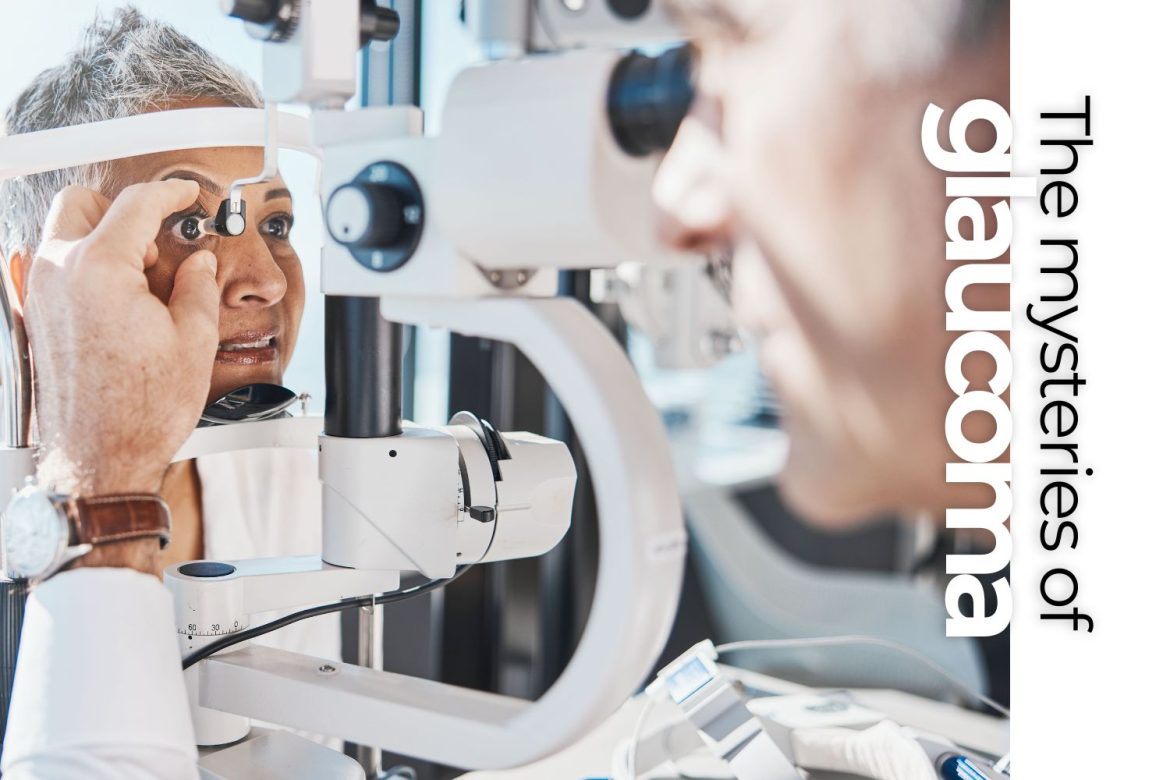
Tonometry measures the pressure inside your eye using an instrument called a tonometer. It is one of the primary tests for diagnosing glaucoma and the target for glaucoma treatment.
Your pressure changes throughout the day so you may be asked to have your pressure checked at different times of the day. IOP is usually highest at 2 or 3 a.m., so early treatment may require using drops at night to prevent an overnight increase.
Ophthalmoscopy
The doctor uses a special magnifying device to examine the optic nerve for signs of damage and assess the condition of the optic nerve.
The doctor will look at your cup to disc (c/d) ratio which is a measurement of the inside of the cup versus the size of the disc. A large c/d ratio can indicate glaucoma; however, you can have a large c/d ratio without glaucoma, which is called physiological cupping.
Perimetry (visual field test)
This test maps out your entire field of vision, identifying areas where you have lost peripheral vision. This is the test where you hit the button when you see the lights. This test can be difficult to take, and the results sometimes improve with practice.
Gonioscopy
The doctor uses a special lens to examine the angle where the iris meets the cornea. This is helpful in determining the type of glaucoma, and whether the angle is open or closed.
Pachymetry
This test measures the thickness of your cornea using an ultrasonic wave instrument. Corneal thickness can affect IOP readings and provides additional diagnostic information. Sometimes patients have high IOP readings because their corneas are thick and not because they have glaucoma. Having LASIK thins the cornea and may affect IOP readings.
Optical coherence tomography (OCT)
This imaging test provides a lot of information for diagnosing glaucoma. It captures detailed images of the optic nerve and retinal nerve fiber layer. It is used to detect early signs of optic nerve damage and to monitor glaucoma progression.
Treatments for glaucoma
While there is no cure for glaucoma, treatment can help slow or prevent further vision loss, especially if the disease is found early.
The four main treatments include: medications, laser treatment, surgery, and lifestyle choices.
Medications
Eye drops are the most common initial treatment for glaucoma. Medications such as prostaglandins, beta blockers, alpha agonists, and carbonic anhydrase inhibitors help reduce intraocular pressure. Doctors expect for the medications to drop the IOP by 30%.
Laser treatment
Laser Trabeculoplasty is a procedure used to treat open-angle glaucoma. The laser is used to open the drainage canals, allowing the aqueous humor to drain more effectively.
Laser Iridotomy is a procedure used for angle-closure glaucoma. A laser creates a small hole in the iris to improve the drainage of the aqueous fluid.
Surgery
Trabeculectomy is a surgical procedure where part of the eye’s drainage system is removed to create a new channel for fluid to leave the eye.
Minimally invasive glaucoma surgery (MIGS) is the newest technique in glaucoma treatment. The devices are less invasive and improve fluid drainage with fewer complications and a faster recovery time.
Conclusion
Glaucoma requires early detection and ongoing management to prevent significant vision loss. With advancements in medical treatments and surgical options, most patients with glaucoma can manage their condition and maintain a good quality of life.
If you have a family history of glaucoma or it’s been a while since you’ve had a routine eye exam, it’s time to see an eye care professional to discuss appropriate screening and treatment options.


